National Toxicology Program (NTP) scientists unveiled the Rodent Ultrastructure Atlas, a digital resource that details the intricate subcellular architecture and organization of various tissues as seen through an electron microscope, May 1. The first-of-its-kind atlas offers a baseline reference for scientists seeking to identify disease- or exposure-induced changes in rodent cells and tissues.
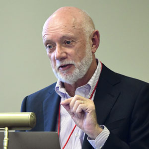
To diagnose such changes, researchers first must know what normal looks like. That is why the Atlas presents normal rodent ultrastructure in detail and in a format that researchers can easily understand, according to Ron Herbert, D.V.M, Ph.D., an NIEHS pathologist and senior scientist who led the team that developed the resource.
“The Atlas, which contains more than 300 high-resolution images supplemented with expert-reviewed descriptions, standardized terminology, and references for further study, will become an essential reference for pathologists worldwide,” added Robert Sills, D.V.M, Ph.D., who leads the Comparative and Molecular Pathology Branch at NIEHS.

A critical scientific tool
Although light microscopy remains the standard for tissue evaluation and disease detection, it cannot provide the subcellular resolution necessary to understand subtle changes, conditions, or abnormalities.
“The view through a light microscope might indicate that cells of an organ are enlarged, but it cannot explain why or how,” Herbert explained. “In contrast, ultrastructure evaluation using electron microscopy can pinpoint effects on specific components of a cell or indicate that a compound has reached its subcellular target.”
The ultrastructure of cells often reflects or correlates with the tissue or organ function. So, understanding normal subcellular morphology is essential to interpret alterations that affect tissue and organ function.

Free, accessible, and intuitively designed
Housed on the NTP website, the Atlas is freely available to the global research community. The digital format — accessible via computer or mobile device — enables users to quickly move between topics and to zoom in for close-up views of the images.
The Atlas offers users an intuitive navigation that organizes information by organ system — such as endocrine, immune, or respiratory — and then individual organs. Each chapter contains images of semi-thin sections, offering general views of tissues and cells, and ultrastructural images to highlight and differentiate subcellular components.
“For the first time, we have put the information all in one place so that researchers can easily go online and quickly find what they want to see,” Herbert said. “No source is as focused, comprehensive, easy to locate, or simple to use.”
A living reference
Although publication of the Atlas marks the culmination of a five-year effort, the project is far from complete.
Plans are underway to add examples of abnormal ultrastructure of disease and disease processes especially related to toxicity and carcinogenicity. Herbert already has solicited images from the global community of toxicologic pathologists and researchers. The new information will be added to the site as it becomes available.
“The beauty of an online resource is its flexibility,” Herbert said, “As new findings emerge, we can rapidly update or expand the content, ensuring it remains relevant and current.”
(Douglas Murphy, Ph.D., is a technical writer-editor in the NIEHS Office of Communications and Public Liaison.)